Veterinary imaging guide
Veterinary Digital Radiography: DR System Basics and Implementation
Veterinary digital radiography converts an X-ray exposure into a digital image that can be reviewed, adjusted, stored, and shared from a workstation. A successful DR project depends on more than the detector: the generator, table, positioning workflow, software, image archive, network, training, and service plan must work as one system.
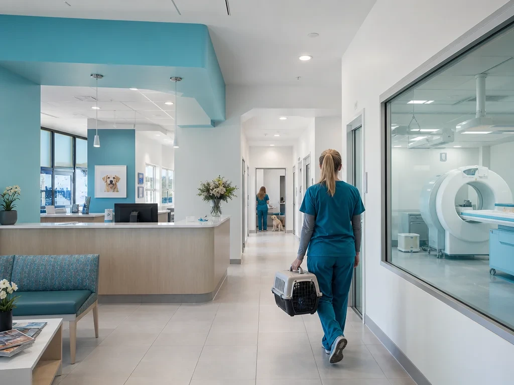
Veterinary Digital Radiography
DR is a film-free X-ray workflow. The X-ray generator creates the exposure, a digital detector captures the transmitted signal, and acquisition software displays the image on a computer. The result is still a radiograph, so positioning, collimation, exposure selection, and radiation protection remain essential.
The British Veterinary Nursing Association's review of radiography systems distinguishes conventional film-screen radiography, computed radiography (CR), and direct digital radiography (DR). It explains that DR sends the image from the detector to the workstation without the separate plate-reading step used by CR. See the verified BVNA radiography review for the underlying system definitions.
The practical takeaway
Do not evaluate a detector in isolation. Start with patient mix, expected study types, room constraints, network and archive requirements, staff workflow, and service access. Then specify the detector and generator around that operating model.
For a practice building a broader imaging service, compare the DR project with the complete veterinary imaging equipment plan. A new facility should also place the room inside its overall veterinary clinic equipment layout and use a starting-a-clinic checklist to catch non-imaging dependencies such as patient handling, power, networking, and recovery space.
DR System Basics
A veterinary digital radiography system is a chain of connected components. Image quality and throughput can suffer when one part is underspecified or poorly integrated.
| Component | Role | Questions to resolve before purchase |
|---|---|---|
| X-ray generator and tube | Produces and shapes the exposure. | Will the available output and controls support the species, body regions, and table or field workflow you expect? |
| Detector | Captures the image signal and transfers it to the workstation. | What detector size, wired or wireless connection, protection, battery workflow, and warranty fit daily use? |
| Table, stand, and positioning tools | Align the patient, detector, and beam while limiting motion and repeats. | Can staff position routine and difficult patients without unsafe manual holding? |
| Acquisition software | Selects studies, previews images, applies processing, and records annotations. | Are veterinary body-part protocols, species fields, user controls, and export functions clear? |
| DICOM and PACS workflow | Standardizes image files and supports archiving, retrieval, and sharing. | Who owns storage, backups, remote access, migration, and integration with the practice system? |
| Service and quality control | Keeps hardware, calibration, software, and staff procedures reliable. | What installation, acceptance testing, training, preventive maintenance, and response process is included? |
A dedicated veterinary X-ray DR system page can help you translate these questions into an equipment brief. For a complete hospital, place that brief inside a pet hospital medical equipment plan instead of treating radiography as a stand-alone room.
Benefits over Film
Digital radiography removes wet chemical processing and physical film storage from the image workflow. DR also removes the separate reader step used by CR, so the image can appear at the workstation soon after exposure. Digital tools support zoom, measurements, annotations, brightness and contrast adjustments, and easier image distribution.
| Decision factor | Film-screen | CR | DR |
|---|---|---|---|
| Image capture | Film in a cassette | Reusable imaging plate | Digital detector |
| Processing step | Chemical processing | Plate is carried to and scanned by a reader | Image transfers from detector to workstation |
| Review tools | Physical film and lightbox | Digital viewing tools | Digital viewing tools with a shorter acquisition path |
| Storage and sharing | Physical archive and manual transport | DICOM/PACS capable | DICOM/PACS capable |
| Main workflow tradeoff | More handling and consumable steps | Digital benefits, but reader handling remains | Fastest path from exposure to review, with greater detector dependence |
Digital post-processing does not correct poor positioning, motion, missing anatomy, or an unsuitable exposure technique. It can also make an image look acceptable across a broad exposure range, so teams still need protocol review and repeat-image monitoring. The goal is diagnostic consistency, not simply a brighter screen.
Where DR Fits in a Veterinary Imaging Service
DR is strong for fast projection imaging of bone, thorax, abdomen, and many emergency presentations. It does not replace every modality. A sound equipment plan selects each tool for the clinical question and the practice's patient mix.
General radiography and dental imaging
General DR and intraoral dental imaging solve different positioning and detail requirements. Plan the veterinary dental imaging suite as a related but distinct workflow, including its own sensor, software, patient positioning, and procedure space.
Imaging works best when connected to the rest of the diagnostic pathway. In-house testing may pair radiographs with veterinary blood analysis, fecal analysis, or urine sediment examination, depending on the presenting problem.
The wider point-of-care veterinary diagnostics workflow should define how results are reviewed together. A separate animal blood analyzer decision needs the same attention to training, sample handling, reporting and referral limits.
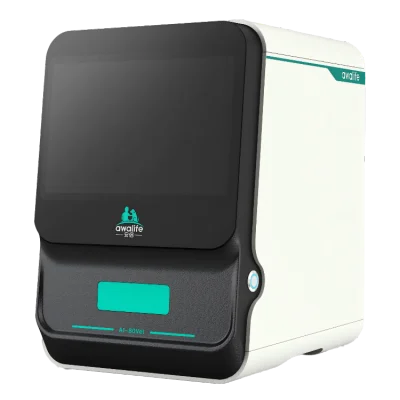
AI-80Vet
A compact option for automated analysis of multiple biological sample types.
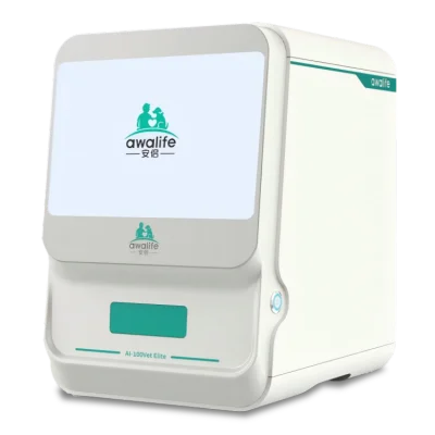
AI-100Vet Elite
An AI image-recognition and microfluidic platform for multi-sample morphology workflows.
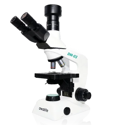
DM-03 digital microscope
PC-connected live imaging, annotation, screenshots, and template-based reporting.
The veterinary microscopy imaging workflow and a properly planned veterinary laboratory equipment area can complement radiography when cytology, morphology, or sample-based evidence is needed.
Implementation Guide
Use the following sequence to turn a DR purchase into a functioning clinical service. The sequence is intentionally vendor-neutral and avoids unsupported price assumptions.
-
1. Define the clinical workload
Document species, body regions, emergency use, dental needs, daily study volume, mobile requirements, and referral expectations. An animal hospital equipment plan may need different room capacity and redundancy than a small practice.
-
2. Survey the room and supporting infrastructure
Confirm power, structural layout, shielding review, controlled access, table clearance, patient transfer, network coverage, workstation placement, and data backup. Include day-to-day veterinary practice supplies and positioning aids in the room plan rather than treating them as an afterthought.
-
3. Specify the connected workflow
Map patient registration, worklist entry, exposure, image review, annotation, interpretation, archive, referral sharing, and record retention. Ask how DICOM, PACS, backups, user permissions, and future data migration are handled.
-
4. Evaluate suppliers beyond the detector
Request a written scope for installation, acceptance checks, training, software updates, preventive maintenance, spare parts, remote support, and escalation. Distributors comparing portfolios can use the veterinary medical supply company framework, while larger procurement programs may need veterinary medical equipment bulk sourcing or veterinary supplies wholesale coordination.
If local market coverage matters, define responsibilities through a formal veterinary equipment distribution cooperation process. Vetsplex's knowledge base supports an integrated sourcing role across medical imaging, diagnostics, surgery, and hospitalization, but it does not provide a published DR model specification or public price on which to base a product claim.
-
5. Train, test, and monitor
Train staff on positioning, exposure protocols, detector handling, image quality, radiation protection, software, backups, and downtime procedures. Track repeats and rejected images by reason so technique and workflow problems can be corrected.
-
6. Connect DR to the full care environment
Coordinate imaging with exam rooms, laboratory testing, surgery, hospitalization, recovery, and referral workflows. Vetsplex plans pet hospital design and equipment projects across these systems. Even supporting tools such as a stethoscope for veterinarians should be assigned by clinical workflow, not purchased as an isolated catalog item.
For wider procurement context, compare animal medical equipment, veterinary haematology analyzers, and the veterinary medical products catalog. Published project cases should only be used when they contain enough detail to match your facility, patient mix, and implementation scope.
Prepare a DR implementation brief
Share your target patient mix, room constraints, preferred workflow, archive needs, installation location, and service expectations. Vetsplex can discuss the DR requirement within a broader veterinary imaging or hospital equipment solution without inventing a generic price or one-size-fits-all specification.
Frequently Asked Questions
What is the difference between CR and DR in veterinary radiography?
CR records the exposure on an imaging plate that must be carried to a reader and scanned. DR sends the image from a digital detector to the workstation without that separate plate-reading step.
Can a digital detector be added to an existing X-ray generator?
Sometimes, but compatibility must be assessed as a system. A qualified supplier should review generator condition and controls, detector integration, table or stand geometry, software, network, acceptance testing, and local regulatory requirements before proposing a retrofit.
Does digital radiography eliminate repeat exposures?
No. Digital processing can improve display, but it cannot restore missing anatomy, remove motion, or fix poor positioning. Staff training, suitable protocols, detector care, and repeat-image review remain necessary.
What should a veterinary practice ask about image storage?
Ask about DICOM support, PACS ownership, storage capacity, backup and recovery, remote access, user permissions, referral sharing, cybersecurity responsibilities, retention, and the process for exporting or migrating the archive.